Infrared spectroscopy for life science
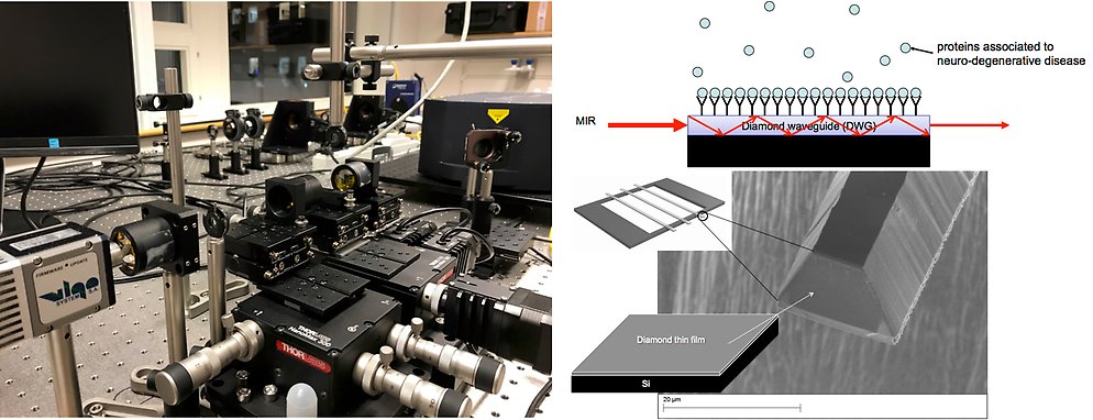
(left) Photograph showing the optical setup of the biosensor, consisting of a broadly tunable QCL, IR-optics and IR-detectors (top right) Schematic showing how a specific protein from a complex mixture can be captured on the diamond waveguide surface. (bottom right) Scanning electron microscope picture of a diamond waveguide end face which is cleaved by focused ion beam milling.
We have during the last years been working on the microfabrication of diamond waveguides.
Life science
Infrared spectroscopy for life science
We have during the last years been working on the microfabrication of diamond waveguides. The diamond waveguides together with a broadly tunable laser quantum cascade laser (QCL), emitting from 5.5 µm up to 11 µm, are the two key elements in our newly developed label free biosensor based on vibrational spectroscopy. When coupling infrared (IR)-light through the waveguide an evanescent wave will be created at the waveguide surface which will interact with the analyte. Because of the reduced thickness when using waveguides, in combination with using QCL lasers as a light source, an ultra sensitive sensor is expected (compared to ATR-IR spectroscopy, which is an existing technology). Moreover, our waveguide can be functionalized which can be used to bind antibodies to the sensor surface.
The waveguides are fabricated by standard lithographic techniques followed by inductively coupled plasma etching of diamond. We are also performing simulations of the light propagation in the different types of waveguides.
In our lab we have a complete optical setup of the biosensor, consisting of a broadly tunable QCL, IR-optics (lenses etc.) for coupling the light into the diamond waveguide, an infrared camera to visualize the IR-beam profile when exiting the diamond waveguide, and a sensitive MCT-detector.
We have demonstrated first measurements on different types of analytes (e.g. isopropanol) at low concentrations (ng) to demonstrate the sensitivity of the sensor.
Currently we are working on analyzing different forms of the protein alpha-synuclein, which is relevant in understanding the mechanism behind Parkinson’s disease. Recently, we used ATR-IR spectroscopy to analyze the secondary structure of different alpha-synuclein aggregates. Interestingly, it seems to be possible to see the difference in the IR-spectra between the native state and the neurotoxic misfolded state of the protein. Our sensitive diamond waveguide biosensor will be used to analyze the secondary structure of alpha-synuclein at biologically relevant concentrations. Future work includes the functionalization of the diamond waveguide sensor surface to be able to fish out alpha-synuclein from cerebrospinal fluid, with the ultimate goal to detect Parkinson’s disease at an early stage.
News
Article demonstrating our newly developed ultra-sensitive biosensor. Next step is to analyze biological relevant samples.
Collaboration
This project is performed in collaboration with Prof. Håkan Engqvist and Prof. Wei Xia (Materials in Medicine, Uppsala University), Prof. Lars Österlund (Solid State Physics, Uppsala University), Dr. Per Ola Andersson (Swedish Defence Agency), Prof. Fredrik Nikolajeff (Luleå University) and Assoc. Prof. Joakim Bergström (Molecular Geriatrics, Uppsala University).