Dmrt3 cells and Renshaw cells in spinal motor circuits
Spinal cord interneurons coordinate locomotion and the integration with descending signals from motor centres in the brain. The direct link between circuit activity in the locomotor CPG and motor output makes it well suited for elucidating the assembly and identity of the participating neurons.
One of the first inhibitory interneuron characterized is the Renshaw cell, which is located in the ventral horn of the spinal cord and mediate recurrent inhibition of alpha motor neurons. While the Renshaw cell act of recurrent inhibition is known since a long time, the exact function of these cells in motor control is yet to be established.
Renshaw cells have been shown to be rhythmically active during fictive locomotion, both in cat and mouse and are likely involved in control of the speed of locomotion. However, an unambiguous role of Renshaw cells is yet to be found. Interestingly, we have found that Renshaw cells are positioned to provide disinhibition (Wootz et al., JCN, 2013, 521:1449-1469) and have been shown to be spontaneously active at rest, firing at a resting rate of 7 Hz (Perry et al., Eur J Neurosci. 2015, 41:889-900).
Neurons affecting locomotion
We previously found that the Dmrt3 gene has a major effect on the pattern of locomotion in mice and horses (Andersson et al., Nature 2012, 488:642-6). This positioned the Dmrt3 gene in a pivotal role for configuring the spinal circuits controlling stride in vertebrates.
In a follow-up study, we establish that Dmrt3 expressing neurons is, at least in part, responsible for the observed effects in the locomotor network (Perry et al., J. Neuroscience, 2019, 39:1771-1782). Moreover, we found that Dmrt3 neurons are inhibitory, connect directly to motor neurons and receive inhibitory, excitatory and modulatory inputs.
We could also show, using retrograde rabies tracing in lumbar Dmrt3 neurons, that these neurons receive inputs from motor-related brain areas and proprioceptive primary sensory neurons in mice (Vieillard J et al., J Comp Neurol. 2023, 531:5-24). This places them in a strategic position to provide sensory-motor control and directly connect to motor neurons, similar to the classical reflex pathways in the spinal cord.
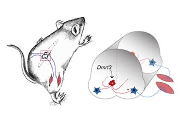
Schematic of the Dmrt3 neurons (red) and relation to motor neurons (blue) in the mouse spinal cord.
Dmrt3 in locomotor control
In collaboration with the Boije group (also in the research programme), we have performed studies that shed light on the role of Dmrt3 in locomotor control. The single-cell transcriptomic analysis revealed distinct subtypes and novel subpopulations of spinal Dmrt3 neurons in zebrafish and mice, providing a better understanding of their complexity and diversity (Iglesias González et al., Front Cell Neurosci. 2021, 15:781197). The behavioural characterization of dmrt3a mutant zebrafish further supports the importance of Dmrt3 in regulating acceleration and locomotor control in vertebrates (Del Pozo et al., eNeuro, 2020, 7:ENEURO.0047-20.2020.
We continue to define the function of neurons coordinating the locomotor circuits in the mammalian spinal cord with a focus on the functional roles of Dmrt3 interneurons and Renshaw cells.