Professor Emeriti - retired professors
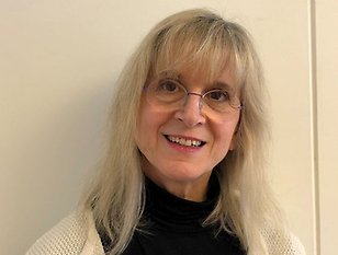
Pia Ek - From regulation to evaluation
I started to study chemistry at Uppsala University 1965, deviating into higher courses in biochemistry, one of which was for Jerker Porath, exploring different coupling techniques to make affinity gels, that later became big business. I collaborated with him again in 1992 separating phosphoproteins on IMAC-columns. As one of the first students, I read the Biomedical Science Program for the possibilities to come even closer to human natural science. After my study I choose the Department of Medical Chemistry (Uppsala university) for graduate studies, since I had most knowledge in this area.
In 1973 I was introduced in Lorentz Engström´s research group. He worked at the time mainly with regulation by protein kinase A phosphorylation, that initially became my research area. The collaboration with Lorentz Engström continued in the field of protein A and C phosphorylation. As a EU Copernicus coordinator for a research project in regulatory phosphorylation in plants, I met and collaborated with several inspiring reseachers, some of which were Ulf Ragnarsson (Uppsala university, Grażyna Muszyńska (Polish Academy of Sciences Warszawa) and Jaak Järv (Tartu university). Around 2000, Örjan Zetterqvist and I decided to collaborate in the field of regulatory phosphorylation. Örjan Zetterqvist had detected a phosphohistidine dephosphorylating activity and since phosphohistidine is difficult to work with and few did it, it was a good choice for us, since we had to do a lot of the practical work ourselves. Our latest publication shows that phospholysine is a substrate for the phosphohistidine phosphatase as well. Due to my teaching I had the possibility to learn all that was to know about biochemical methods, a knowledge that also are reflected in my publications.
Memorable publications are those concerning phosphorylation of pyruvate kinase, since they were work that I did as a graduate student and they were original at the time. And the collaboration with Örjan Zetterqvist with his profound knowledge in phosphohistidine and his perfectionism was an inspiring time and an enriching experience.
I was told that I was the last in a row of researchers working with phosphoproteins at the department. First out was Gunnar Ågren, followed by Lorentz Engström, Örjan Zetterqvist and last me.
After my retirement 2013 I changed research area. As I had a son born 1978 with severe intellectual and developemental disability (IDD) I was forced to learn about the conditions for persons with IDD in the community and health care and wanted to contribute in order to improve their health. Therefore a collaboration with Päivi Adolfsson (Uppsala university) started in the field of health related to nutrition.
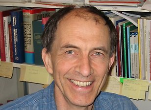
Erik Fries - In and out of cells
With no particular career plan but with an interest in physics, I started my university studies in 1967 with mathematics followed by physics. However, the physics courses were not as interesting as I had hoped so I continued with general chemistry, physical chemistry and biochemistry. I then wanted to do more pratical work and was accepted as a PhD student at the Department of Biochemistry.
My project concerned the release of proteins from membranes by detergentmolecules. This project led me to a cooperation with a group at the European Molecular Biology Laboratory (EMBL) in Heidelberg that was studying the interaction of different detergents with a membrane-containing virus and the last year of my PhD studies I spent in their lab. After defending my thesis in May 1977, I was allowed to stay one more year as a postdoc at the EMBL and then began studying the initial interaction of the virus with its host cell. My analysis of the pH-dependence of the binding of the virus to different cells led us to the discovery of the process by which this and some other membrane bounded viruses infect cells: through internal pH-dependent fusion of viral and endosomal membranes.
In October 1978 I went to the laboratory of James Rothman at Stanford University for a second postdoc period. Rothman wanted to elucidate the molecular mechanisms underlying the vesicular transport of proteins from the endoplasmic reticulum (ER) to the cell surface. To achieve this goal he wanted me to reconstitute the transfer of a newly synthesized protein destined for the plasma membrane from the ER to the Golgi complex in a cell homogenate. However, this approach turned out to be unsuccessful, but I managed to obtain an inter-Golgi transfer in my cell free extract. Unexpectedly, this reaction became the basis for Rothman´s further work which earned him the Nobel Prize in 2014.
In November 1980 I returned to Uppsala to work on protein secretion with Prof Per Peterson at the Department of Cell Research. Here I was introduced to the use of primary cells in the form of hepatocytes isolated from rat liver. After about two years I got a position as an assistant professor at IMBIM. Here I continued my work on proteins secreted by hepatocytes, in particular how these proteins are modified during their transport to the cell surface. One of the proteins I studied was the chondroitin-sulphate containing protein bikunin, which, in a complicated way, is involved in the formation of the extracellular matrix. In 1990 I became associate professor and in 2000 full professor.
Paraskevi Heldin - my privileged life as scientist
My scientific life started in 1981, when I started as a graduate student in the laboratory of the eminent professor Lorentz Engström, at the Department of Medical and Physiological Chemistry, studying regulatory phosphorylation of proteins. After my dissertation in 1987, I started to work on hyaluronan biology, as a postdoctoral fellow in the laboratory of professor Torvard Laurent, who worked on the same department. Under Torvard´s enlightened leadership, I became dedicated to the hyaluronan field and particularly to the investigation of the effect of over-expression of hyaluronan in inflammation, cancer and infection.
As was known in the 1980-ies, an overproduction of hyaluronan occurred in blood and tissues under rapid tissue remodeling and infection, where a plethora of growth factors and cytokines are released. At that time, there was a growing interest both in clinical work on hyaluronan, and in basic research to elucidate the molecular mechanisms underlying its excess production during disease. In 1983, CD44, which first was described as the receptor leading to the extravasation of inflamed lymphocytes into inflammatory foci, was interestingly found to be the principal cell surface receptor for hyaluronan. This finding united immunology and extracellular matrix research, and it was possible to understand why one of the physiological functions of hyaluronan was to capture circulating cells, such as lymphocytes and neutrophils, and lead them to inflamed sites.
My subsequent career involved employment as Head of the Matrix Biology Group at the Ludwig Institute for Cancer Research, Uppsala, where world leading research on growth factor signaling was performed. In this inspiring and productive environment, our studies ontargeting of hyaluronan-CD44 signalling in cancer and infection revealed a correlation between tumor progression and growth-factor-mediated hyaluronan synthesis and CD44 over-activity.
In 2017, I became a guest professor at the Department of Medical Biochemistry and Microbiology (IMBIM) and continued our research on hyaluronan-CD44 signaling. In this nice environment, we demonstrated that perturbation of hyaluronan signaling contributes to the progression of diseases, such as aggressive breast cancer, glioblastoma, Dengue virus infection and COVID-19 infection. I formally retired in 2022, but am still working on the elucidation of the underlying mechanisms of CD44 signaling and on the development of ways to inhibit hyaluronan-CD44 signaling.
%20(3)%20birgitta.jpg)
Birgitta Heyman - 45 years of antibody feedback regulation
Antibodies can regulate the antibody response against the antigen they bind to. This phenomenon is called antibody feedback regulation and can be both positive, leading to more than 100-fold enhancement, or negative, resulting in almost complete suppression. My research has been aimed at understanding the molecular mechanisms behind these regulatory circuits.
In 1978 I started as a PhD student with professor Hans Wigzell as supervisor at the Department of Immunology at Uppsala University. By then I had realized that working with patients was not my cup of tea and that research would be more fun. Hans presented me with a paper by Niels Jerne and Claudia Henry from 1968 showing that IgM anti-SRBC (sheep red blood cells), passively administered together with SRBC to mice, enhanced the antibody response against SRBC. In the same situation, IgG anti-SRBC almost completely suppressed the antibody response. My PhD project was to elucidate the mechanisms behind these dramatic immunoregulatory effects of IgM and IgG and this area has continued to fascinate me throughout my career.
With the exception of a postdoc period at Scripps Clinic and Research Foundation, La Jolla, California in William Weigle's lab (1983-84) and a visiting professorship at Harvard, Boston in Michael Carroll's lab (2001-02), I have been employed by Uppsala University, albeit at several different departments: Department of Immunology, Department of Medical Chemistry, Department of Pathology, Department of Genetics and Pathology (where I became professor of Experimental Pathology in 1999 and professor of Experimental Immunology in 2002). In 2008, my group moved to Department of Medical Biochemistry and Microbiology where I am now professor emerita.
IgG-mediated suppression. The suppressive effect of IgG takes place whether or not the IgG antibodies can activate complement or bind to Fc-receptors. This, together with many other experimental findings, suggests that suppression is in fact caused by mere binding of IgG to the antigen, thereby hiding it from recognition by the immune system. IgG-mediated suppression is used clinically to prevent haemolytic disease of the newborn, which can occur in Rhesus positive fetuses carried by Rhesus negative women. Whether the mechanism above applies also to human Rhesus prophylaxis is currently a matter of debate but I find it unlikely that they should be unrelated phenomena.
IgM-mediated enhancement. The other part of my PhD project was to explain why IgM antibodies enhanced the antibody response. After returning from Scripps and having started my own lab, we could show that mutant monoclonal IgM antibodies which had lost their ability to activate complement also lost their enhancing ability. Much later, in collaboration with Michael Carroll, we constructed a knock-in mouse (Cm13) with the same point mutation as the mutant IgM antibody. This mouse had a slightly reduced antibody response showing that endogenous IgM, via complement, indeed plays a role in upregulating antibody responses. However, the reduction was far from as pronounced as that seen in complement deficient mice and thus cannot fully explain the role of complement in antibody responses. The most likely explanation for IgM-mediated enhancement is that antigen-IgM-complement immune complexes are efficiently transported to areas in the spleen where immune cells interact with antigen to initiate antibody responses.
IgE-mediated enhancement. In 1993, we found that IgE, passively administered together with protein antigens, could enhance antibody responses, sometimes more than a 100-fold. In a series of papers, the last one published in 2016, this multi-step process was investigated. It turns out that IgE-antigen complexes after immunization are rapidly captured by B-cells circulating in the blood and expressing an IgE-receptor, CD23. Within 30 minutes, the IgE-complexed antigen (but not uncomplexed antigen) can be found in the B cell areas of the spleen and is then transferred to a subtype of dendritic cells. These endocytose and present peptides to specific CD4+ T helper cells which increase dramatically in numbers during the first 3 days. The efficient help given by these T cells to antigen specific B cells most likely explains the enhanced antibody responses observed.
IgG-mediated enhancement. A fourth branch of our feedback regulation studies concerns the ability of IgG antibodies, administered together with protein antigens, to enhance antibody responses. One of the murine IgG subclasses, IgG3, executes this through the same mechanism as IgM, i. e. antigen-IgG3-complement complexes are being formed and transported to the "right" areas for immunostimulation in the spleen. Interestingly, the other IgG subclasses do not rely on complement for their enhancing effects but instead use Fc-receptors for IgG. Most likely, the antigen-IgG complexes bind to Fc-receptors on dendritic cells which endocytose them, and present peptides to CD4+ T helper cells, subsequently offering efficient help to antigen specific B cells.
In summary, feedback regulatory effects of antibodies are often caused by direct interference with the antigen. Antigen can be hidden, it can be captured for transport to optimal areas for immune cell stimulation, or it can be endocytosed and presented efficiently to T helper cells. We have found little evidence for direct B cell regulation through inhibitory IgG Fc-receptors or stimulatory complement receptors expressed on the B cells themselves.
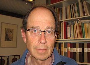
Ulf Lindahl - A personal voyage through the proteoglycan field
Proteoglycans (PGs) are proteins substituted with sulfated polysaccharide (glycosaminoglycan, GAG) chains. My selection of the PG research area occurred by sheer accident. During the course in medical biochemistry, part of the medical curriculum (1959, at the old department on Dag Hammarskjölds väg, opposite to the hospital), the class was subdivided into seminar groups that were arbitrarily assigned to senior teachers.
Lennart Rodén had me run a few GAG samples on Sephadex columns and then departed for University of Chicago, where I subsequently spent the first two years of my graduate training elucidating the structure of the GAG-protein linkage region.
Upon my return to Uppsala, Torvard Laurent took over as supervisor until my graduation in 1966. I remain indebted to Lennart and Torvard for their guidance, support and friendship.
My subsequent career involved employment as professor in medical and physiological chemistry, first at the veterinary faculty at SLU (1973-91) then at Uppsala University (now IMBIM, 1991-2005). When asked why I retired at age 65 rather then the optional 67, I noted that although I had appreciated my profession in general I felt done with certain tasks, such as marking written examinations. Other aspects of undergraduate teaching I quite enjoyed, and it has been rewarding to follow the curriculum develop toward its current problem-oriented state. I recall lecturing medical students on atomic orbital theory!
My scientific work, along with a large number of coworkers, remained focussed on PGs, in particular on their heparin and heparan sulfate GAG chains. In the course of this work emphasis was gradually shifted from structural carbohydrate chemistry to GAG metabolism (biosynthesis and degradation of GAG chains), functional properties and GAG pathophysiology. We still aim at defining the mechanisms of biosynthetic regulation behind the generation of tissue-specific heparan sulfate chains, an ever fascinating problem. A limited number of other groups elsewhere have pursued similar lines of research, realizing the vital roles of PGs (and their GAG-chain ”business ends”) in development, homeostasis and disease. Yet the PG field has remained relatively small; in particular, I am surprised at the relative scarcity of scientists devoting their attention primarily to the GAG chains. It have enjoyed my friendship with several colleagues within this community, including some competitors.
I formally retired ten years ago. Many former graduate students, postdocs, guest scientists and research associates have since showed up around our kitchen table. Some still remain active in the lab. I would like to especially thank the three senior scientists, Lena Kjellén, Jin-ping Li and Dorothe Spillmann, for many years of most enjoyable collaboration, and for their friendly smiles when I still turn up at the lab. Finally, I note that although the department has grown tremendously since the old days, IMBIM remains a pleasant and inspiring scene for scientific pursuit.
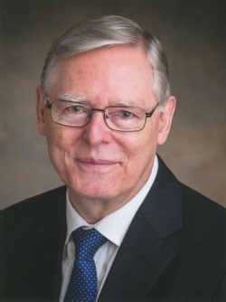
Örjan Zetterqvist - My scientific life
My scientific life began in 1961 at the Department of Medical Chemistry, with a PhD project that aimed at searching for protein‑bound phosphohistidine in mammalian tissues. This kind of histidine modification was unknown at that time, but a year later Paul D. Boyer et al. at the University of Minnesota reported the isolation of phosphohistidine from bovine liver mitochondria. Eventually they identified this phosphoamino acid as the 3‑phosphohistidine isomer, using methods that became important in my own work.
With Lorentz Engström as my tutor my PhD research continued. There is an inherent difficulty in working with phosphohistidine due to the acid-lability of its N‑P‑bond. Nevertheless, my studies of the rapid formation of protein-bound phosphohistidine in rat liver cytosol led to the first detection of protein-bound 1‑phosphohistidine in biological material. In later studies from the laboratory we could identify this phosphohistidine isomer as a true intermediate of the nucleoside diphosphate kinase reaction by using a rapid‑mixing technique that permitted the study of reactions with a half-life of a few milliseconds. We also detected protein‑bound 3‑phosphohistidine in the liver cytosol, and a few years later I could show that most of this isomer originated from rapidly autophosphorylated ATP-citrate lyase.
The rapid‑mixing equipment, which I had designed, was also used in studies of the intermediary phosphorylation of sodium-potassium ATPase, a project further developed by Sven Mårdh.
In the early 1970s Lorentz Engström, together with most of his research group, switched to the study of proteins phosphorylated by cyclic AMP-dependent protein kinase (PKA). This was how the well-known regulatory phosphorylation of liver pyruvate kinase was discovered. This phosphorylation occurs as a consequence of the hormonal stimulation of the gluconeogenesis in liver during starvation. Further studies in which I participated led to the first identification of a consensus amino acid sequence around the phosphorylatable serine of a PKA‑substrate. These studies, and several that followed, benefited greatly from collaboration with Ulf Ragnarsson at the Department of Biochemistry on the synthesis and characterization of a large number of phosphorylatable peptides.
For a protein phosphorylation to be regulatory it must be reversible, which requires the action of a protein phosphatase. This aspect was studied in collaboration with Vincent Titanji, then a PhD student at my laboratory. My long‑standing contact with him and some of his students to support their studies on tropical parasites in Cameroon led to an extensive correspondence that was recently filed in the IMBIM archive.
Around 1980 I found it worthwhile to switch focus and I chose to investigate the possible existence of enzymes that could modify phosphorylated sites by other processes than dephosphorylation. This search was initiated by adding a set of 32P-phosphorylated peptides of varied length to rat liver extracts, and led to the discovery of tripeptidyl peptidase II. The initial characterization of the enzyme was performed with Ros-Mari Bålöw. The project was then taken over by Birgitta Tomkinson, who developed it extensively and successfully.
During the first years of the 1990s I made an attempt with Johan Rosén to look for additional modifications of phosphopeptides, which led to the rediscovery of prolyl endopeptidase and the discovery of its preference for a phosphorylated substrate.
After arranging a Wenner‑Gren International Symposium on protein phosphorylation in September 1997 in honour of Lorentz Engström and as I was approaching my formal retirement in 2002, I ventured to return to the phosphohistidine field. The studies on the histidine phosphorylation of the bacterial phosphoenolpyruvate‑dependent phosphotransferase system and two‑component systems had become extensive at that time, while the progress of the studies on phosphohistidine in mammalian cells had been slower. By joint efforts with Gunilla Pettersson and by use of a phosphohistidine‑containing peptide as a probe, we succeeded in discovering a 14‑kDa phosphohistidine phosphatase (PHPT1) in porcine liver. The further characterization of this enzyme was performed through an invaluable collaboration with Pia Ek, who became the principal investigator of the project. The successful expression of the recombinant human enzyme was made possible by support from Jin-Ping Li. PHPT1 has proved to be a valuable tool in phosphohistidine research. Our first report on the enzyme was published in 2002. A brief review can be found in the introduction of our most recent paper (1).
In this context it should be mentioned that discovery of the same enzyme was independently reported in 2002 by Susanne Klumpp and her group in Münster. Sadly, Dr Klumpp died in 2009, which was a great loss for all of us investigating this enzyme.
I hope to be able to conclude my scientific activity by following the research on phosphohistidine in mammalian cells for some time to come. There now seems to be a gradually increasing interest in this field, due to its possible relevance in understanding part of the metabolism of cancer cells. In addition, recent adaptations of phosphoproteomic methods for detection of protein-bound phosphohistidine give hope for the future.