Transmission Electron Microscopy
Transmission Electron Microscopy (TEM) enables you is to aquire high-resolution images of cellular ultrastructures and small particulates. A beam of electrons is transmitted through an ultrathin specimen, usually contrasted with heavy metals, and is scattered in correlation to the specimen mass-density. Electromagnetic lenses focus and magnify the transmitted electrons on to a CMOS or CCD camera, which forms the final image. At the BioVis TEM Node we are ready to help you with your scientific questions, whether you work at the University of Uppsala, other Swedish academic institutions or in the industry. The services we offer are conventional sample preparation, negative staining, immunogold labeling and imaging. We have experience in a wide range of different types of samples and protocols. In case you are interested in more advanced techniques such as single particle analysis (cryo EM) or 3D imaging (FIB-SEM) these instruments can be found at BMC and Ångström respectively.
If you want to discuss a TEM project or have further questions, please contact BioVis´ EM staff member Monika Hodik and Karin Staxäng. Price settings are found under Start here menue.
Electron Microscope TEM, Tecnai G2
TEM Tecnai G2
The BioVis EM node offers its customers a Tecnai G2 BioTwin Spirit transmission microscope from FEI/Thermo Fisher. We encourage our customers to sit independently and aquire images to ”get to know” their samples and the advantages/limitations of the microscope. We provide an introduction to the basic operation for imaging. Please note that we charge for Introdcution and using the instrument.
Summary of TEM Tecnai G2
- 80-120kV (Lab6 filament)
- Resolution: 0.2 nm minimum
- Magnification: up to 300.000x
- Camera: Gatan Orius SC200 CCD, 2048x2048 px, 30fps
Contact EM BioVis staff for more information.
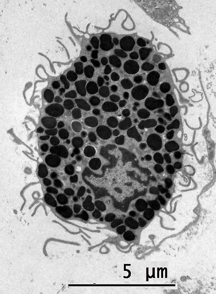
Image:
TEM image of a mast cell. Magnification: 4200X
Embedding Service (Resin)
Conventional sample preparation is commonly used to preserve ultrastructural morpholgy. The sample preparation includes steps of perfusing, fixating and dehydrating the sample, embedding it into Epoxy resin, followed by ultrathin sectioning and contrasting.
There are several challenges regarding the sample preparation for biological TEM. The sample must be fixed properly to avoid ultrastructural changes due to degradation, it must be dehydrated to withstand the vacuum inside instrument and cut to ultrathin sections to allow electrons to be transmitted. In addition, since biological material mainly consist of elements with a low atomic numbers (H,O,C), heavy metals need to be added to increase the contrast. All steps impact the final result and must be carried out with great carefulness.
Summary of Resin embedding protocol
- Secondary fixation in Osmium Tetraoxid
- Dehydration in increasing concentrations of Ethanol
- Propylen Oxide
- Infiltration of Epoxy resin (Epon, LR White or Spurr)
- Polymerization by heat
- Semi-thin sections stained with Toluidine Blue
- Ultrathin sections (50-60 nm)
- Slices are collected on grids
- Contrasting with Uranyl Acetate and Reynold’s lead citrate
- Imaging at 80kV (Resolution up to 1.0 nm)
Contact EM BioVis staff for more detail regarding sample preparation.
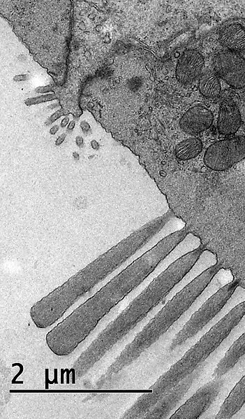
Image: TEM image of an short inner hair cell in the inner ear of an Cuban crocodile. Magnification: 9900X
Kindly provided by Helge Rask-Andersen, Pubcare, Uppsala University Hao Li et al., Front. Cell Dev. Biol, 2022
Immunogold Labeling Service
Immunogold labeling is a technique that allows to visualize (the position) of proteins in samples like tissue, cells, suspension. To detect the protein of choice, target-specific antibodies are utilized - the so called Primary Antibodies. Then Secondary Antibodies are used, to target the primary Antibodies. The Secondary antibodies are conjugated with colloidal gold particles at specific sizes, which can be visualized easily in the TEM.
It is possible to do pre-immunogold labeling, in which the antibodies are added befor resin embedding or post-immunogold labeling, in which the antibodies are added after resin embedding, i.e. directly on ultrathin sections. It is also possible to combine immunogold labeling with negative staining.
Final sample preparation includes different steps to allow for preservation of the ultrastructure and production of ultrafine slices, needed for imaging. These steps different will interfere with antigenicity. Hence, the signal in this Immunogold labing is generally lower compared to other immunolabeling techniques due to those harsher embedding procedures. Therefore we encourage our customers to first try e.g. immunofluorescence on their samples, to ensure that the target protein is present and can be detected.
BioVis can provide its customers with goat anti-mouse and goat anti-rabbit secondary antibodies. All other antibodies will be charged extra. In case small/ultrasmall gold particles are used (<2nm), we provide silver enhancement to increase the size and visability of the gold particles.
Contact EM BioVis staff for more detail regarding sample preparation.
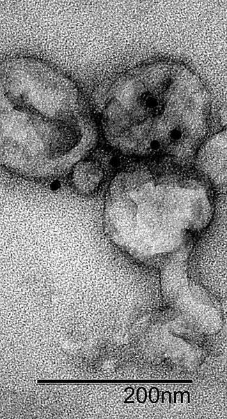
Image: TEM image of immunogold labeled negatively stained exosomes. Magnification 87 000X
Kindly provided by Ana Rosa Saez Ibanez.
Negative Staining Service
Negative staining. This rapid and easy method is used to visualize small particulates like proteins, exosomes, lipids, virus, filaments and more. It is often used to validate the presence of the particle of interest. In addition more high-resolution techniques (cryo) are preceded by negative staining. It is possible to combine immunogold labeling and negative staining.
A drop of the sample in solution is put on a supportive grid. Excess of the solution is removed by a filter paper followed by addition of a contrasting agent (usually uranyl acetate). After removing the contrasting agent by a filter paper, the grid with the sampleis left to air dry before imaging in the Electron Microscope. The sample will be imaged at 80-200kV resulting in an resolution of app. 2nm at its best, depending on the sample.
Contact EM BioVis staff for more detail regarding sample preparation.
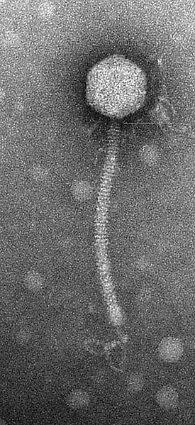
Image: Negative stain TEM image of bacteriophage. Magnification: 105 000X, Kindly provided by Marcelo Gutierrez-Valverde, IMBIM, Uppsala University.
Featured Image
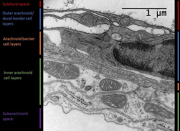
The transmission electron microscopy (TEM) image shows a cross section of the adult mouse arachnoid mater, the middle meningeal membrane, and its surrounding spaces. The cellular complexity of this structure was recently revealed (Pietilä et al, Neuron 2023). The combination of single-cell RNA sequencing, high-resolution fluorescence microscopy and TEM shows that the arachnoid consists of four different fibroblast-like cell types organised into distinct layers, along with extracellular matrix components some of which are also seen in the TEM image.
Courtesy of Christer Betsholtz.
Pietilä R, Del Gaudio F, He L, Vázquez-Liébanas E, Vanlandewijck M, Muhl L, Mocci G, Bjørnholm KD, Lindblad C, Fletcher-Sandersjöö A, Svensson M, Thelin EP, Liu J, van Voorden AJ, Torres M, Antila S, Xin L, Karlström H, Storm-Mathisen J, Bergersen LH, Moggio A, Hansson EM, Ulvmar MH, Nilsson P, Mäkinen T, Andaloussi Mäe M, Alitalo K, Proulx ST, Engelhardt B, McDonald DM, Lendahl U, Andrae J, Betsholtz C.Neuron. 2023 Dec 6;111(23):3745-3764.e7. doi: 10.1016/j.neuron.2023.09.002. Epub 2023 Sep 29.PMID: 37776854