Light Microscopy
Imaging is a balance between speed and resolution and contrast. Different specimens are better suited to different microscopes. Accordingly we provide microscopes to match a wide variety of specimens from whole animals to individual cells, with different resolutions from organs to superresolution as well as different imaging speeds.
For Fluorescence Microscopy, Slide Scanner and Confocal Microscopy (LSM700) Contact Jeremy Adler .
For STED Microscopy, Two Photon Microscopy and Confocal Microscopy (Stellaris) Contact Matyas Molnar.
In order to use any of our instruments - you need to either book a full service or attend to our Instrument introductions. Application to instrument introduction can be found under Introductions. Price settings are found under Start here menue.
Instruments are bookable over our booking system.
Widefield Microscopy (BF&FL)
Zeiss AxioImager
The AxioImager is an upright microscope for fluorescence and transmitted light widefield microscopy. It is suitable for thinner samples where 3D information is not priority.
Summary of ZEISS AxioImager
- Upright
- Widefield Epifluorescence, Brightfield
- Objectives 5x/0.15, 10x/0.45, 20x/0.8, 40x/1.3 oil, 63x/1.4 oil
- Excitation Source: HXP 120C lamp
- Emission filters: DAPI, GFP, CY3, A647
- Cameras: (i) AxioCamMRC(RGB); (ii) AxioCamMRm (monochrome)
- Motorized stage XYZ
- Tiling and stitching
The LSM700 can also be used a a widefield Microscope utilizing its Cameras. More information under Confocal Microscopy menue
Contact jeremy.adler@igp.uu.se for more information.
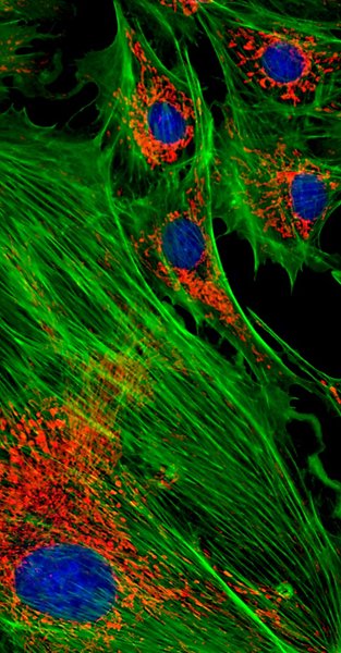
Image: Cells with stained nuclei (blue), green actin Cytoskeleton, and red Mitochondria
Slide Scanner (BF&FL)
Zeiss Axio Scan.Z1 BF FL
Create a recipe for finding the tissue, focusing and then imaging up to 100 slides automatically – press start and walk away.
Summary of SlideScanner
- Upright
- Widefield Epifluorescence, Brightfield
- Objectives 5x/0.25, 10x/0.5, 20x/0.8, 40x/0.95
- Excitation Source: LEDs 385, 475, 555, 630, white (transmission)
- Emission filters: 430-470, 500-555, 570-640, 665-759, multi band pass
- Cameras: (i) ORCA (monochrome); (ii) Hitachi (RGB)
- Motorized stage – tiling
- Z series
Contact jeremy.adler@igp.uu.se for more information.
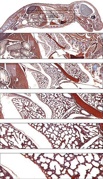
Image: Whole slide of whole Mouse section, including different inserts to show acchieved resolution using 10x objective. Inserts show increasing magnification. Original image: 40kx20k pixels
Confocal Microscopy
Zeiss LSM700
The Zeiss LSM700 is a state of the art inverted confocal microscope with 4 lasers 2 fluorescence and one transmitted light detector. With both oil, water and air objectives it suits different specimens and resolutions. Specimens like cells, zebra fish and organs can be imaged on slides, in petri dishes or on multiwell plates. The LSM700 can also be used a a widefield Microscope utilizing its Cameras.
Summary of LSM700
- Inverted
- Lasers: 405/488/555/633
- Two PMT detectors for simultaneous detection, transmitted light PMT
- Objectives: 10x/0.5, 20x/0.8, 40x/0.95, 25x/ multi immersion, 63x/1.2 water, 63x/1.4 oil
- Motorized scanning stage
- Tiling
- Photobleaching
- Positions
- Images in XYZ, time
- Incubator, temperature control
- Widefield microscopy application using Cameras
Download the LSM700 manual Pdf, 2 MB.
Contact jeremy.adler@igp.uu.se for more information.
BioVis gratefully acknowledges the financial support from MedFarm FISK and IGP department which has allowed the purchase of the LSM700 (2014).
Leica Stellaris 5
The Stellaris 5 is a versatile workhorse confocal microscope from basic fluorescence imaging to advanced microscopy experiments. An instrument serving most user needs and especially designed to a wide variety of users from beginners to experienced. Equipped with white light laser for flexible excitation and Tau time resolved imaging for better signal characterization.
Summary of Stellaris 5
- Inverted
- Lasers (405, plus white light laser 485-790nm)
- Three Hyd detectors for simultaneous detection
- Free choice of emission bandwith for detection
- Objectives: 10x/0.4; 20x/0.75; 63x/1.4 oil
- Motorized scanning stage
- Fast overview images
- Tiling and stitching
- Images in XYZ, time
- Tau, time resolved imaging of stained and unstained samples
Download the Leica Stellaris 5 manual Pdf, 2 MB.
Download the Leica Stellaris 5 Tau guide Pdf, 3 MB.
Video tutorials on how to use the LAS X software can be found under Leica Microsystems Tutorials on Youtube: video 1, video 2.
Contact matyas.molnar@igp.uu.se for more information.
BioVis gratefully acknowledges the financial support from MedFarm FISK and IGP department which has allowed the purchase of the Leica Stellaris 5 (2023).
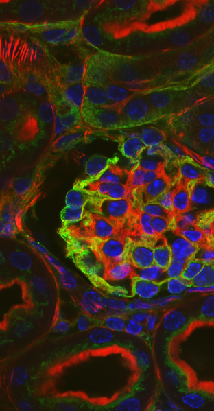
Image: Kidney glomeruli
STED Superresolution Microscopy
Abberior Stedycon
To take a next step in resolution, STED offers an enhanced resolution in the XY direction, up to 30-40nm. The Z resolution is unchanged, similar as for confocal imaging. The higer resolution is achieved by using a torus shape infrared depletion laser that can turn off special fluorophores in a selected XY area. Thus, for this technique special dyes are needed (se below) and for the best result users should follow good sample preparation routines for microscopy.
Summary of Stedycon
- Upright
- Excitation lasers: 405/488/561/640; and STED depletion:775nm
- Dye examples for confocal imaging: DAPI; GFP/AF488; Cy3/AF568; Cy5/AF647.
- Dye examples for STED: AF594/Abberior STAR 580-600; AF647/Abberior STAR RED.
- Objectives: 100x/1.45 oil, WD: 0.13mm (for STED); 10x/0.3 air (for overview)
Download the STEDYCON manual Pdf, 5 MB.
Contact matyas.molnar@igp.uu.se for more information.
BioVis gratefully acknowledges the financial support from MedFarm FISK and IGP department which has allowed the purchase of the Abberior STEDYCON (2020).
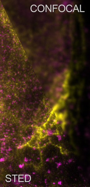
Image: Comparison of STED and Confocal imaging with a sample of fixed cells
Multiphoton Microscopy
Leica SP8 DIVE
In many cases we need to look deeper in biological samples to see a bigger picture of the specimen and the surrounding biology. Multiphoton microscopy (synonyms: two-photon microscopy, nonlinear microscopy) is a tool using short-pulsed high power infrared laser for penetrating deeper in biological samples as a normal confocal laser could. This tool can be very useful when deeper 3D imaging in necessary with limited photobleaching. The multiphoton system is a scanning imaging technique; therefore its limitation is speed. For 3D rendering, Imaris is available at BioVis.
Summary of Leica SP8 DIVE
- Upright
- Motorized stage for small animal imaging
- Multiphoton laser: 680 - 1300nm tunable line; 1045nm fixed line
- Objectives water, n=1.33 (motorized corr. collar): HC IRAPO 25x/1.0; HC FLUOTAR L 16x/0.8
- Objective clarity, n=1.45 (motorized corr. collar): HC FLUOTAR L 25x/1.00
- Tiling and stitching
- Vario beam expander
Download the Leica SP8 multiphoton manual Pdf, 4 MB.
Contact matyas.molnar@igp.uu.se for more information.
BioVis gratefully acknowledges the financial support from MedFarm FISK, IGP department and the Beijer Foundation which has allowed the purchase of the Leica SP8 DIVE (2019).
Image: Second Harmonic Generation of skeletal muscle