Heteroresistance & Antibiotic Susceptibility Testing
Overall theme of the projects revolve around studying cell-to-cell variation in growth rates and how they are related to antibiotic resistance of various kinds. We are developing various tools to identify genetic reasons for complex phenotypes at single cell level and do ultra-fast diagnostics of complex bacterial infections. These tools involve employing advanced microfabrication techniques, machine learning (cell detection and tracking), optics (fast imaging and tweezing single cells) and software development (for real-time analysis).
a. (Main-Project) Heteroresistance detection and mechanism identification
Heteroresistance has been recently identified as a major caveat to the antibiotic susceptibility testing(AST) using minimum inhibitory concentration(MIC) measurements[1]. The presence of a small resistant subpopulation of cells in the main population of susceptible cells can’t be detected on the fast ASTs[2] due to their low frequencies (1 in 103-107 ) and their exact mechanisms are relatively unstable and hard to study. In this project we develop microfluidic chips that can be used for large scale (~100,000) single cell phenotype and growth measurements and optical tweezing technique[3] to pick single cells for heteroresistance mechanism identification by single-cell sequencing.
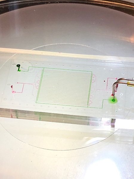
Functioning microfluidics chip
b. (Secondary Project) Fast multi-species antibiotic-susceptibility testing
In this project, we are developing ultra-fast protocols for doing normal AST based on growth rates and doing species identification using fluorescent probes[4]. The key challenge is to disentangle species information from the growth curves and discover the differential impact of antibiotics during a diagnostic test.
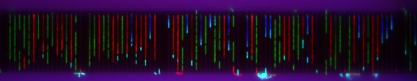
Microfluidics chip with different bacteria in the channels.
Related published research
- Nicoloff, H., Hjort, K., Levin, B.R. et al. The high prevalence of antibiotic heteroresistance in pathogenic bacteria is mainly caused by gene amplification. Nat Microbiol 4, 504–514 (2019)
- Özden Baltekin, Alexis Boucharin, Eva Tano, Dan I. Andersson, Johan Elf. Antibiotic susceptibility testing in less than 30 min using direct single-cell imaging. Proceedings of the National Academy of Sciences Aug 2017, 114 (34) 9170-9175; DOI: 10.1073/pnas.1708558114
- Luro, S., Potvin-Trottier, L., Okumus, B. et al. Isolating live cells after high-throughput, long-term, time-lapse microscopy. Nat Methods 17, 93–100 (2020).
- Amann, R., Fuchs, B. Single-cell identification in microbial communities by improved fluorescence in situhybridization techniques. Nat Rev Microbiol 6, 339–348 (2008).
- Yichao Yang, Kalpana Gupta, Kamil L. Ekinci. All-electrical monitoring of bacterial antibiotic susceptibility in a microfluidic device. Proceedings of the National Academy of Sciences May 2020, 117 (20) 10639-10644;