Molecular imaging and radionuclide techniques
Molecular imaging in biomedical research
Molecular imaging is the concept of noninvasive studies of biochemical and physiological processes in living animals and man. It is increasingly important in clinical research and diagnostics, as well as in development of new pharmaceuticals. Two very important technologies are positron emission tomography (PET) and magnetic resonanace imaging (MRI) and recently the fusion between PET and MRI has been realised. This fused technology is known as PET-MRI. In addition to PET there is single photon emission computed tomography (SPECT) which also can be combined with CT.
PET-MRI, a state-of-the art fusion of two technologies
Conventional PET-CT scanners co-register the functional PET images with Computed Tomography (CT) images obtained just prior the PET examination. However, PET images can also be manually co-registered with Magnetic Resonance Imaging (MRI) acquired at a different time point than the PET scan. This is preferable since MRI provides markedly improved contrast between soft tissues and is especially useful for studies of the brain. Additionally, functional MRI (fMRI) enables non-invasive in vivo assessment of physiological parameters.
This has prompted the development of integrated PET-MRI scanners. MRI provides structural imaging of the body by exploiting the behavior of hydrogen nuclei in the presence of strong magnetic fields. As it does not use ionized radiation (as opposed to CT) PET/MRI yields a markedly reduced radiation dose, which enables safer examinations of new patient groups or healthy subjects. In addition, the PET and MRI scanning can be performed simultaneously instead of sequentially which improves image alignment and reduces artifacts due to movement.
Preclinical PET-MRI
Preclinical small animal PET-MRI has improved resolution for both functional (<1 mm) and structural imaging (<100 μm) compared to most current preclinical instruments. In vivo biodistribution, drug pharmacokinetics and pharmacodynamics, therapeutical effects, tissue function and blood-brain barrier penetration can thus be studied at an unparalleled depth and resolution.
The preclinical PET-MRI scanner also offers the potential of several functional in vivo measurements of functional parameters crucial for evaluation of drug or therapy effects. These include tissue perfusion by both PET (H2150) and MRI (contrast enhancement), regional oxygen consumption (oxygen-15 and BOLD, respectively) and molecular imaging (MRI spectroscopy).
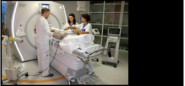
Human PET-MRI scanner
Translational PET-MRI in collaboration with Uppsala PET centre Uppsala University hospital
In October 23, 2014, Sweden’s first human PET-MRI equipment was inaugurated at Uppsala University Hospital. The nationally available human equipment funded by a grant from the Swedish Research Council will for example be used for whole body examinations to for instance detect tumours and evaluate therapies.
The non-invasiveness of PET-MRI investigations allows rapid translation between the preclinical and the clinical setting. Only minute amounts of radiolabelled compound (far below pharmacological doses) are required to be administered for clinical imaging due to the high sensitivity of modern PET scanners (the principle of “microdosing”). This means that the risk of adverse effects is greatly diminished.
More information about ongoing radiology research at the Department of Surgical Sciences.
Radiochemistry
At PPP we offer expertise in radiolabelling and radiochemistry. Our well equipped laboratory with shielded fume hoods makes it possible to radiolabel a wide array of small molecules and macromolecules for molecular imaging. We work with a range of different PET and SPECT radionuclides (see below). For purification and analysis we have two HPLC systems.
Do you have a molecule in your project you want to label? Please get in contact with us as early as possible in your development process so we can discuss your needs and how to incorporate radiolabelling chemistry strategies.
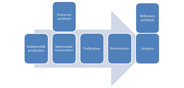
Radiochemisty work flow
Do you have a molecule in your project you want to label? Please get in contact with us as early as possible in your development process so we can discuss your needs and how to incorporate radiolabelling chemistry strategies.
Radionuclide |
| Imaging method | Half-life t1/2 | Comment |
|---|---|---|---|---|
Carbon-11 | 11C | PET | 20.4 min | Cyclotron production at UUH |
Fluorine-18 | 18F | PET | 109 min | Cyclotron production at UUH |
Cobalt-57 | 57Co | SPECT | 271 d | Commercially available |
Gallium-66 | 66Ga | PET | 9.49 h | Cyclotron production |
Gallium-67 | 67Ga | SPECT | 78.3 h | Commercially available |
Gallium-68 | 68Ga | PET | 68 min | In house generator |
Zirconium-89 | 89Zr | PET | 78 h | Commercially available |
Technetium 99m | 99mTc | SPECT | 6 h | Generator produced at UUH |
Indium-111 | 111In | SPECT | 67 h | Commercially available |
Iodine-123 | 123I | SPECT | 13.2 h | Commercially available |
Iodine-124 | 124I | PET | 4.18 d | Commercially available |
Iodine-125 | 125I | SPECT | 59.4 d | Commercially available |
Iodine-131 | 131I | SPECT | 8 d | Commercially available |
Lutetium-177 | 177Lu | SPECT | 6.6 d | Commercially available |
Rehnium-188 | 188Re | SPECT | 16.9 h | Commercially available |
Carbon-11, t1/2 20.4 min, PET
Carbon-11, t1/2 20.4 min, PET
Uppsala PET centre at the University Hospital produces range of 11C-labelled PET tracers in the routine production such as 11C-methionine, 11C-hydroxytryptophane, 11C-PIB. In addition, in principle any tracer synthesis can be setup for preclinical and clinical trials provided the precursor and reference compund are available in reasonable amounts. Uppsala PET Centre and Uppsala University is internationally well recognised for the innovation and development of [11C]methyl iodide and [11C]carbon monoxide labelling chemistry.
Fluorine-18, t1/2 109 min, PET
Fluorine-18 is very versatile for labelling of small molecules and in used in many routine PET tracers such as 18F-FDG, 18F-FMISO and 18F-FLT. Such routine traces can sometimes be obtained form the Uppsala PET Centre. A range of routine tracers are also available from PPP by the use of the casstte based GE FastLab2 synthesizer. At PPP we also develop own labelling protocols for cusoms molecules with 18F. The conditions required for fluorine carbon bond formation is often harsch so the labelling is often performed in multiple steps and with the use of protecting groups. Therefore the chemical route of labelling requires careful design.
Gallium-68, t1/2 68 min, PET
Gallium-68 is one of the most verstile radionuclides for labelling of small peptides. We have our own preclinical 68Ge/68Ga-generator that can be eluted 2-3 times daily. Uppsala PET Centre have facilities to produce 68Ga-tracers under cGMP conditions for clinical reserach.
Gallium-66, t1/2 ___min, PET
Gallium-66 has a longer half-life than gallium-68 and cab be produced by a medical cyclotron.
Zirconium-89, t1/2 78 h, PET
Larger molcules such as proteins often have slower kinetics in vivo and therefore a radionuclide with longer halflife is required. Typically the radionuclde inocorporation can be performed using Desferrioxamine B (DFO) bioconjugates at room temperature and pH 6.8-7.2.
Indium-111, t1/2 67 h, SPECT
Peptides, protein and other macromolecules functionalised with a chelator such as DTPA, DOTA, NOTA or NODAGA can be radiolabelled with standard protocols.
Radio-iodine
Similar protocols can be used for all radio iodine, however, individual optimisation might be required due to different contaminants during the radionuclide production. In our laboratory we extensively use iodine-125 to radiolabel proteins for initial SPECT studies, and iodine-124 for PET-studies.
Lutetium-177, t1/2 6.6 d, SPECT
Lutetium-177, t1/2 6.6 d, SPECT
Used with chelators such as DOTA and DTPA. Used for both imaging and radiotherapy studies.
In vitro methods
In vitro autoradiography
We use a cryostate for sectioning of organs or tumours into thin slices which can be applied to a piece of glass.
Ex vivo autoradiography
The animal is sacrificed and the organs are immediately frozen and sliced with the cryostate.
Radiometabolite studies
The amonunt of intact tracer is an important parameter when performing kinetic modelling. That can be obtained by analysing samples of blood, urine and homogenized organ extracts by radio-HPLC.
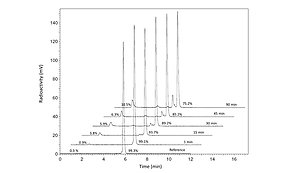
HPLC radiometabolite analysis