Methodology

Methodology
Methodology and infrastructure
Photoelectron spectroscopy (PES)
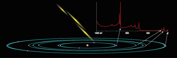
Photoelectron spectroscopy (PES) is based on the photoelectric effect and involves measurement of the kinetic energy of electrons that are emitted from a material as a result of their interaction with incident x-ray light. The energy of these photoelectrons gives information on the electronic structure and molecular composition in the sample.
PES is an element specific technique since each element has its own characteristic electronic binding energy distribution of its core level electrons. Smaller adjustment of this binding energy distribution, e.g. chemical shifts, occur due to the interaction with surrounding atoms. Since also the intensity of the emitted photoelectrons correlates with the density of a specific atom it is possible to determine the composition of a sample.
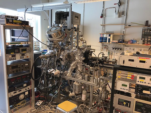
ELEPHANT lab (Ångström laboratory, house 6, floor 0).
SOXPES (Soft X-ray PES) and HAXPES (Hard-X-ray PES)
PES is a surface sensitive technique with a probing depth that can be varied from a few nanometers up to tens of nanometers. The development of synchrotron radiation sources has enabled the use of a wide range of monochromatic photon energies. SOXPES (soft-X-ray photoelectron spectroscopy) and HAXPES (Hard X-ray photoelectron spectroscopy) are two synchrotron-based PES techniques using different photon energy regions, approximately hν < 1500 eV for SOXPES and hν > 2000 eV for HAXPES. The latter is more and more present at synchrotron facilities including beamlines at DESY, BESSY, Diamond and Soleil.
In our research, the modification of the incoming photon energy is a very important tool as it allows us to probe our sample with different probing depth and gives us information over a thickness from a few Ångströms to a few tenths of nanometers. It also allows us to investigate the character or hybridization of the valence states since the cross-section for photoemission depend on the photon energy then depend on the valence level composition.

Panorama of Bessy II.
Ambient Pressure Photoelectron spectroscopy (APPES)
Photoelectron spectroscopy measurements commonly require ultra-high vacuum conditions, which cause volatile substances, e.g. a liquid electrolyte, to be pumped away from the surface of the sample. Recent development of photoelectron spectroscopy instrumentation allows for measurements at higher pressures, so-called ambient pressure photoelectron spectroscopy (APPES), under which pressures a liquid can be stabilized. The division is currently going a step further with the development of the methodology and the instruments to combine the surface analysis and the battery cycling to achieve APPES measurements while the battery is charged or discharged, i.e. going from classical ex-situ post-mortem to in-operando PES measurements on battery materials.
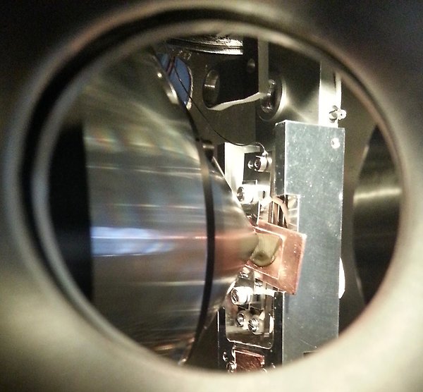
APPES measurement on Si electrode with electrolyte drop.
Resonant inelastic X-ray scattering (RIXS) and X-ray absorption spectroscopy (XAS)
RIXS and XAS are two techniques that provide complementary information about the electronic structure of materials. These are excellent techniques for studying samples in situ and in ambient pressure environments, e.g. Li ion batteries which contain liquid electrolytes and operando catalytic water-splitting studies. XAS usually is typically used to probe the empty states close above the Fermi level of a material. RIXS can either be used to determine neutral excitations in the range between a few meV to several eV or the simply occupied states below the Fermi level of a material. Both techniques have the advantage of providing orbital specific and x-ray polarization sensitive information. A specific new possibility will be the VERITAS beamline at MAXIV.
RIXS has recently experienced a rapid development attracting an ever-increasing number of users and resulting in a quickly rising number of high-profile publications. This development has been made possible by the availability of brilliant synchrotron radiation at new facilities around the world. We perform XAS and RIXS at leading synchrotron facilities and beamlines worldwide, e.g. ADRESS@SLS and BL8@ALS. The X-ray Photon Science division also has a decades-long track record of developing high resolution grating spectrometers for cutting edge research using RIXS. A new advanced x-ray spectrometer has been developed to match the unique capabilities and ultra-high resolution of VERITAS at MAX IV laboratory in Lund and is currently being implemented there.
We have developed, and are continuously improving, sample manipulation platforms containing reaction cells for in situ and operando x-ray studies. These are sample cells that allow flow-through of liquids and gases past an x-ray transparent membrane window. If desired, a solid film may be deposited on the membrane that interacts with the fluid. Examples include the study of atmospheric corrosion, control of water content in gels, heterogeneous catalysis and Li-ion battery cathodes in contact with a liquid electrolyte.
Energy material research at HELIOS
Functionality of the modern devices used in the energy applications often depends on the behaviour of the non-equilibrium charge carriers (e.g. photoexcited electron-hole pairs or conduction electrons). In order to study this, the pump-probe time resolved PES (tr-PES) can be used. One of the beamlines in our in-house HELIOS HHG laboratory is specifically designed for the time-resolved pump-probe spectroscopy and allows to perform angle-resolved pump-probe photoelectron spectroscopy measurements with the overall time resolution better than 50 fs.
HELIOS HHG source produces ultra-short photon pulses (< 35 fs) and covers the energy range from 20 eV to 72 eV, which is ideal to study the valence states of molecules/solids and shallow core levels. The pump photons with the wavelength of 800 nm and 400 nm are currently available. The experimental station is equipped with the modern Angular resolved Time-Of-Flight spectrometer (ArTOF 2) from Scienta Omicron. The high electron transmission of the ArTOF spectrometer (compared to the standard hemispherical electron analyser) allows to minimize the number of photons per pulse required to achieve the good statistics in the photoemission spectra. Moreover, the high angular acceptance of the spectrometer allows to perform “single-shot” Angular Resolved PES (ARPES) measurements while covering the reasonably big fraction of the k-space in both kx and ky directions.
First experiments [1,2] have demonstrated high efficiency of the setup and its advantages compared to the analogous instruments available at the synchrotron radiation facilities. Among them are the very good time resolution (tens of -fs in compare to tens of -ps at the synchrotron), low signal to noise ratio (due to the absence of the higher diffraction orders in the HHG light), stability of the pump-probe synchronization (the same laser is used to produce pump and probe photons).
[1] U. B. Cappel et al., Phys. Chem. Chem. Phys. 18, 21921 (2016)
[2] J. Terschlüsen, Doctoral Thesis, Uppsala University, 2016 (full-text available)
Energy material research at UBjL
At the Infrastructure Uppsala-Berlin joint Laboratory our division is engaged in the build up and running of two beamlines. Both beamlines employ time-of-flight electron spectrometers that have larger transmission at the same resolution as a conventional hemispherical electron analyser. The beamlines exploit this in different ways: the LowDosePES (a) beamline is situated at a dipole source which, together with an x-ray chopper, allow for x-ray radiation sensitive materials to be studied; the second beamline, CoESCA, have two ArTOF spectrometers with the goal of using coincidence spectroscopies. The LowDosePES beamline is equipped with a picosecond laser system that together with the time-structure of the synchrotron X-rays allow us to use pump-probe techniques to study excited electronic states and phase transitions on a picosecond timescale (b). Some examples of our activities is to follow how electrons and atoms move upon visible illumination in materials such as perovskites and polymers.
(a) Erika Giangrisostomi, et al. Low Dose Photoelectron Spectroscopy at BESSY II: Electronic structure of matter in its native state.
Journal of Electron Spectroscopy and Related Phenomena (2017)
(b) Ute B. Cappel, et al. Partially Reversible Photoinduced Chemical Changes in a Mixed-Ion Perovskite Material for Solar Cells.
ACS applied materials & interfaces, 9.40: 34970-34978 (2017)
Contact
- Programme Professor Condensed Matter Physics of Energy Materials
- Håkan Rensmo
- Head of Division X-ray Photon Sciences
- Nicusor Timneanu